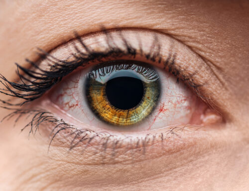
While the eyelids may be taken for granted, they are a delicate and complex part of the body that provides a range of functions. These include protecting the eyes from the environment, blinking to spread the tear film across the eye’s surface, producing the necessary oils for tears to function properly through the meibomian glands, and cosmesis. In this article, we’ll discuss some of these important eyelid functions.
The Eyelid’s Anatomy: Structure and Appearance
When we look at the eyelid from an anatomical viewpoint, it appears to just be a thin fold of skin that covers and protects the eye from foreign debris and dust. However, this is a bit deceptive, as the eyelid is actually quite complicated, and is made up of many structures. Dr. Matthew Shirley of Shirley Eye Care was able to lend us his expertise about the complex structure of the eyelids. Let’s briefly dive into these structures.
1. The Skin & Subcutaneous Tissue
The skin of your eyelid is extremely thin, measuring less than 1 mm in thickness. If you run your finger towards the corner of your eye, near your nose, you’ll notice that this portion of skin is a lot smoother and oilier. This is due to the large number of sebaceous glands in the area, which produce an oily substance called sebum. Its job is to coat, moisturize, and protect the skin.
Just beneath the skin is a layer of tissue called subcutaneous tissue (a connective tissue), also known as hypodermis or superficial fascia. It contains all manner of cells, including elastin fibers, collagen, fibrous bands, nerves, lymphatic vessels, blood vessels, sweat glands, hair follicles and more.
In terms of subcutaneous fat, there is very little in the skin around the eye. There is none where the medial and lateral palpebral ligaments attach, as these connect to the tarsal plate (more about this below).
2. The Orbicularis Oculi Muscle
The orbicularis oculi muscle is what allows you to close your eyelids, and make the facial expressions you do. Every single time this muscle moves, the skin that lies on top of it, also moves. This muscle facilitates blinking, winking, and forcibly closing your eyelid. It has 3 clearly defined sections called the orbital orbicularis, the palpebral orbicularis, and the lacrimal orbicularis.
3. The Submuscular Areolar Tissue
This is loose, connective tissue that forms a dividing plane to separate the eyelid into a front (anterior) and back (posterior) portion. It sits beneath the orbicularis oculi muscle and is what helps develop the upper eyelid crease. It is also responsible for the fullness of the outer upper eyelid (orbicularis oculi fat), and the rounding out of the lower eyelid (suborbicularis oculi fat).
4. The Fibrous Layer
There are several structures that make up this layer.
a. The tarsal plates are a dense fibrous tissue that helps your eyelids maintain both their shape, and their structural integrity. There are two types of tarsal plates, the superior tarsus, and the inferior tarsus, both of which are attached through the medial and lateral palpebral ligament. The superior tarsus takes the form of a crescent, and measures around 10 mm vertically, while the inferior tarsus measures about 3.5-5 mm in height. Both of these come into contact with the eyeball’s conjunctiva (see below for more information).
The tarsal plates also have 25 meibomian glands (upper), and around 20 (lower) around their rim. They produce meibum, an oily substance that prevents your eye’s tear film from evaporating.
b. The medial palpebral ligament is a band of fibrous tissue that holds the inner aspect of the tarsal plate in place.
c. The lateral palpebral ligament is found originating from the tarsal plate, and its role is to help keep structures within the eye stable.
d. The orbital septum is another band of connective tissue that attaches to the border of the orbital bone. It’s role is to join together the lid retractors at the lid margins.
e. The orbicularis retaining ligament attaches the orbicularis oculi muscles to the lower rim of the eye socket.
5. Eyelid Retractors
These are a group of muscles that are responsible for keeping your eyelid elevated. The main muscle that forms a part of this group is called the levator palpebrae superioris (LPS), and forms medial and lateral horns. The lower eyelid retractors form the inferior tarsal muscles, which is what gives the bit of mobility that you do have in your lower eyelids.
6. Fat Pads
In the upper eyelids, you have the aponeurotic fat pad which is found behind the orbital septum, and two other fat pads that are centrally and medially located. The central fat pad is yellow-colored, while the medial is a pale yellow.
In the lower eyelids, you have the same centrally and medially located fat pads, but then there is an additional one that lies in front of the inferior oblique muscle. The role of this one is to elevate the eye.
7. Conjunctiva
This is a smooth, mucous membrane that covers your eye, and lines the inside of your eyelids. It is completely transparent.
8. Other components
Other components of the eyelids include the lacrimal gland (tear production), the lymphatic drainage in the eyelid, and of course the nerve and blood supply vessels found in the connective tissue.
The appearance of the human upper eyelid varies and is dependent on one’s geographic location and culture. In the majority of East Asian and Southeast Asian populations, the epicanthic fold covers the inner corner of the eye. The crease of the eyelid also varies from population to population, usually forming as a singular fold or a double fold.
Age-Related Changes in the Appearance of the Eyelids.
Dermatochalasis is a common age-related change in the upper and lower eyelids, where the loss of elasticity in the skin, weakening of eyelid connective tissues, and gravity causes the skin to become loose. In the upper eyelid, the excess preseptal skin, and sometimes the orbicularis muscle, will droop downwards over the front edge of the eyelid.
With the lower eyelid, you may see bulging orbital fat pockets, but these will rarely impact your vision. In severe cases, individuals with this excess tissue can have their superior visual field blocked up to 50%. Another name for this condition is “baggy eyes”.
How Is Dermatochalasis Fixed?
If the eyes are the “window to the soul”… then the eyelids must be the “window dressings”, or perhaps even better, the “protectors of the soul”. When dermatochalasis gets bad enough, it can impair vision and needs to be fixed. Expert Beverly Hills Plastic Surgeon, Dr. Brian Shafa, explains that the procedure to fix dermatochalasis is called blepharoplasty. It is a surgery that primarily removes the sagging, excess skin. In some individuals, a portion of the orbicularis muscle may be removed, as well as any unneeded fat. This procedure can be performed for visual (medical) reasons, or if there is no obstruction of vision, it can be done for cosmetic reasons (better appearance).
The blepharoplasty treatment is performed under local anesthetic, and ranges from 1-3 hours depending on whether you are getting the upper eyelids, lower eyelids, or both done at the same time. Returning to work takes about 10-14 days, and any type of swelling or bruising in the area fades within 1-3 weeks. The benefit of this surgery is improved vision, but also brighter eyes, and a more youthful appearance.
What Are Eyelid Oil Glands, and How Do They Function?
Along the inner rim of your eyelids, where your eyelashes are found, are meibomian glands, a type of sebaceous gland that produces an oily substance called meibum. This oily layer is the outside of the tear film, which is what keeps your tears from drying up too quickly.
What Happens When the Meibomian Glands Become Blocked?
If your meibomian glands become inflamed (meibomian gland dysfunction), this can cause the glands to become blocked or obstructed by a thick, cloudy-to-yellow opaque substance which is a mixture of oil and wax secretions. An obstruction like this can lead to blepharitis, which is a condition characterized by chronic inflammation of the eyelid, at the base of the eyelashes. The symptoms of blepharitis include soreness, swelling, irritation, burning sensations, crusting, excessive tearing, itchiness, and flakes of oily particles wrapped at the base of your eyelashes.
How Do We Treat Blepharitis?
We got a few tips on how to treat blepharitis from Dr. Kara Shirley of Armstrong Eye Care:
1. Warm Compresses
Using a clean washcloth with warm water can help loosen up an oily residue or flakes that are sticking to the base of your eyelashes and covering/blocking your meibomian glands. By removing the waxy residue in this way, you help keep the nearby glands from clogging.
2. Artificial Tears
Steroid eye drops, or artificial tears can help combat against dry eyes by reducing the amount of redness and swelling within the eyelid. An antibiotic eye drop will help the oil glands work better by reducing infection.
3. LipiFlow (Thermal Pulsation Treatment)
This treatment gently applies pressure and precise heat directly to the upper and lower meibomian glands in your eyelids. The heat melts away any secretions that are blocking your glands, and gets rid of biofilm, bacteria, and mites.
4. Blephex (Electrochemical Lid Margin Debridement)
This is an in-office treatment that cleans the eyelids by removing excess biofilm, bacteria, and bacterial toxins from your eye’s surface glands. It works by gently exfoliating your eyelid margins. It only takes a few minutes to perform, and it helps your eyelids stay symptom-free.
When trying to keep blepharitis symptoms under control, make sure to practice proper eyelid hygiene, by carefully washing out your eyelashes every day. It can be important to get your blepharitis under control before you decide to have cataract surgery.
Talk to your expert cataract surgeon about this before having surgery. You may also want to use antibacterial soap to help keep bacteria growth to a minimum.


